|
| ||||||||||||||||||||||||||||||||||||||||||||||
|
Section: News |
deutsche Version  Print-Version |
|
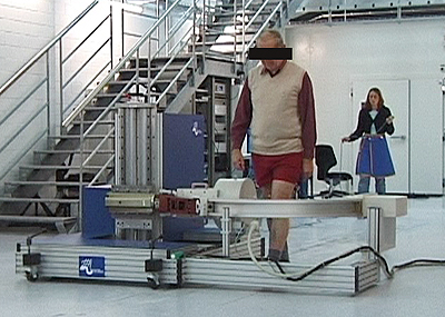
Mobile X-ray unit The knee in motion | |
|
Using a mobile X-ray unit, ETH researchers from the Institute for Biomechanics are able for the first time to X-ray the knee during normal walking. The purpose of the equipment is to help understand how an implanted artificial knee joint behaves during the everyday movement of walking. Christoph Meier Walking is a dynamic process. Nevertheless, up to now the function of artificial knee joints has been analysed using static images of extended and bent knees. However, these were scarcely able to explain why certain patients’ prostheses were painful again and again. This is a big problem, because about one million artificial knees are implanted each year, 40,000 of them in Switzerland. This situation led researchers at the Institute for Biomechanics to analyse the problem in more detail (1). The prosthesis manufacturer Mathys in Bettlach and the medical appliance manufacturer Philips were enthusiastic about the idea and took part in the development. The aim was to develop an instrument with which the prosthesis can be observed during several steps. A sprint buggy If you go into the biomechanical engineers’ test hall on the Hönggerberg today you will see the result of the development. It consists of a platform measuring about 1.5 by 2 metres with a motor on top and four driven wheels underneath. Projecting out on one side is a straight bar that has a wheel and carries a C-shaped arm. At the end of the arm there is a video-fluoroscopy X-ray unit made by the Philips Company. This unit creates the images of the artificial knee by capturing the X-rays on a fluorescent screen. To allow measurements to be made, the test person must stand at a suitable position in the X-ray machine. His knee is then connected to a movement sensor that determines its speed. When the test person runs about 10 metres along the test track, the measuring buggy drives jerkily alongside him. The jerky movement is a consequence of the fact that the knee’s horizontal speed varies greatly during a normal step. The maximum acceleration of the joint during the step is up to 8 m/s^2. Faster than Tom Lüthi? Project leader Hans Gerber’s comments on the consequences: “Our measuring equipment must be able to accelerate as fast as Tom Lüthi.” To give the 450 kilogram apparatus the ability to achieve this, the researchers equipped it with four-wheel drive. The motors reach a maximum torque of about 4000 Nm. However, for successful analysis the X-ray equipment also needs an appropriate resolution. “At present we can photograph 25 images per second,” says Gerber. That is good, but the intention is to improve it even further.
|
The image analysis software already exists. In collaboration with the ETH Institute for Image Processing, the scientists developed an algorithm (2) that enables the three-dimensional conditions to be reconstructed from the two-dimensional images. Regarding this achievement, Hans Gerber says: “It is now possible for the first time to see how the knee behaves during walking. Studies must now be carried out to examine whether there is a relation between the pain and the 3-dimensional situation during walking.” Very promising studies The researchers have already carried out initial studies with knee prosthesis wearers. Although the data is available, Gerber cannot present it because of his obligations to his industrial partners. The researcher indicates only that there are exciting insights. Further studies with the world’s first mobile X-ray unit are also underway. These are intended to establish a relationship between the knee movements and the loading. To do this the researchers are able to measure the floor reaction forces simultaneously with floor panels. Thus a first step towards pain-free walking for all artificial knee joint wearers seems to have been taken already. |
||||||
|
Footnotes:
You can write a feedback to this article or read the existing comments. | |||||||