|
| ||||||||||||||||||||||||||||||||||||||||||||||
|
Section: Science Life |
deutsche Version  Print-Version |
|
New center for animal PET imaging Bright spot provides new insights | |
|
Last Thursday ETH Zurich opened a new centre for PET imaging on its Hönggerberg campus. This is a further stone in the mosaic of Science City, and is to help in the research of various medical disorders and contribute to the further development of this method of diagnostics. Christoph Meier ETH Zurich has a new bright spot – the PET Imaging Center on its Hönggerberg campus. This is not just a figure of speech. The location, equipped with a cyclotron, a synthesis lab and the PET scanner, is bright and shiny, and it will stay that way, because cleanliness is of paramount importance in the work carried out here. Of the centre's 500 square meters, no less than 200 are devoted to clean rooms. The installation, costing a total of 11 million francs, is located underneath the courtyard between the third and fourth "fingers" of the HCI building. At the official opening of the centre, its head, ETH Professor Pius August Schubiger, voiced his conviction that thanks to this new installation ETH would take its place among the world's elite in the area of PET imaging. This had also been made possible by the considerable work already carried out at the Paul-Scherrer Institute. The new PET centre is a collaboration between the PSI, Zurich's University Hospital and ETH Zurich (the latter two already run the Center for Radiopharmaceutical Science(1)).
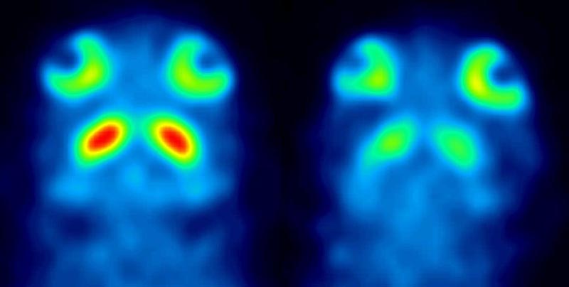
Low-invasive insights For the sake of the assembled media, Schubiger pointed out that PET, which stands for Positron Emissions Tomography, makes it possible to see, not morphological structures, but metabolic processes. This method facilitates the investigation of diverse processes directly inside living organisms. The only surgical intervention necessary is an injection of radioactively labelled molecules. The scientist illustrated the method using the examples of a human tumour and a defect in a mouse brain. In the latter it was apparent that PET provides high-resolution images on the millimetre scale.
|
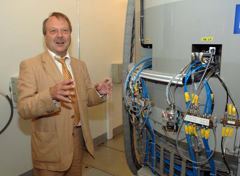
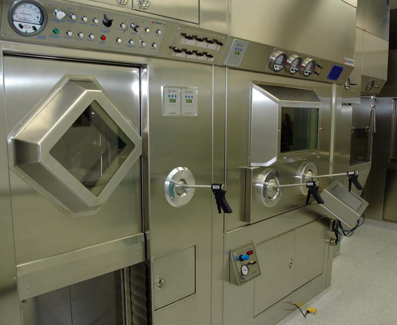
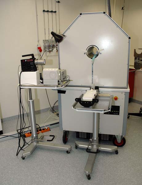
To investigate the processes involved in various disorders – Schubiger also mentioned depression as a possible area of research – substances need to be found which can be radioactively labelled without changing their biological properties. It was in this domain, the development of such substances, that the actual core competency of the center lay, he explained. For its work in this area the center is receiving strong financial support from the pharmaceutical company Schering. This company also made a substantial – but for data protection reasons undisclosed – contribution towards the new centre's installations. From cyclotron to PET scanner In the installation Schubiger explained a full PET process, which, depending on the decay rate of the radioactive nuclides, had to be completed within a few hours. In a first step radionuclides such as C-11 or F-18 are manufactured in the cyclotron, a two-metre-wide vat, weighing 25 tonnes with lots of cables and a complex inner life. The nuclides then move to the synthesis lab, where chemical reaction processes unite them with carrier substances. Of these the best-known is glucose, but for diverse research approaches a whole range of molecules is to be radioactively labelled. In a third step the so-called PET tracers arrive in the room where they are injected into the animal under examination. Thus prepared, the mice or rats are mildly anaesthetised before they are put into the PET scanner. The PET data acquired are then reconstructed to produce its familiar glowing images.
When queried about radiation, Schubiger said that high, dangerous levels can occur in the secure room with the cyclotron. In the trial animal itself, however, the dose is lower than value for normal radioactivity in the environment, i. e. less than 1 to 2 milli-Sievert. |
|||||||||
|
References:
Footnotes:
You can write a feedback to this article or read the existing comments. | ||||||||||