|
| ||||||||||||||||||||||||||||||||||||||||||||||
|
Section: Science Life |
deutsche Version  Print-Version |
|
Pharmaceutical giant Schering to sponsor a cyclotron as well as research for the Imaging Center Millions for "glowing" images | |
|
A year from now an Imaging Center in "Science City" at Hönggerberg will be built, that specialises in preclinical research on animal models. The German pharmaceutical firm Schering will sponsor a cyclotron as well as research projects, which means a total investment of approximately a million euros a year over the next eight years in the new center. By Jakob Lindenmeyer On May26th representatives from Schering (1), together with representatives from Zurich's University Hospital (2) and Professor Pius August Schubiger (3) from ETH Zurich, held an aperitif for the media to inform on the future research co-operation in imaging diagnostics. As part of its "Science City" project ETH is building the Imaging Center at Hönggerberg and setting up the infrastructure into place. For its part, Schering will be investing approximately a million euros a year over the next eight years–half to go towards the running of the center, notably the cyclotron–and half to research.
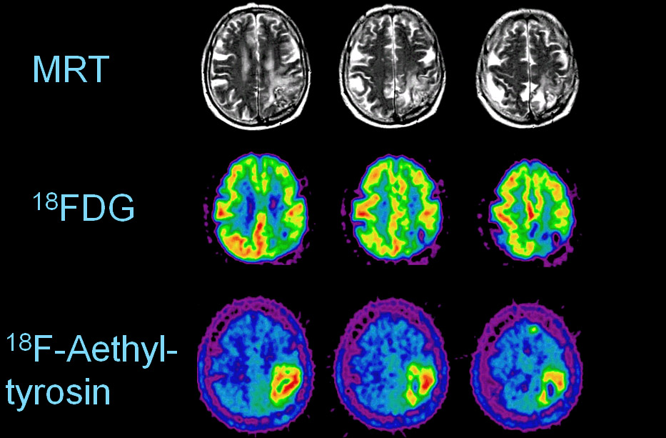
Making cancer and diseases of the brain visible Pius August Schubiger is ETH professor of radiopharmacy and head of the Radiopharmaceutical Centre, a centre run by ETH Zurich, the Paul-Scherrer Institute (PSI) and the University Hospital Zurich. Schubiger will take over as head of the new Imaging Center on the campus Hönggerberg. In his research projects at the Center he will be employing imaging methods, like positron emission tomography (PET, see box), in order to test–more rapidly and more accurately–the suitability of specific of pharmaceuticals in animal models. Until now PET has been used, above all, in the diagnosis of cancer. In future great progress is to be expected in areas of neuro-degenerative diseases, such as Alzheimer's, Parkinson's or multiple sclerosis. The research team working with Schubiger and Simon Ametamey (3) are concentrating on the development of new, specific PET molecules that, when marked with a radioactive atom, become visible with PET. Isotopes, such as C-11, N-13, O-15 and F-18, will be produced by bombarding them with the atoms of accelerated protons from the newly acquired cyclotron. True, the Zurich area already has a cyclotron at its University Hospital but the capacity of this apparatus is fully exploited with clinical examinations. Practically no unoccupied hours exist for ante-clinical research. Schubiger compares the cyclotron at PSI with a ship's motor while delicate, preclinical research, he says, requires rather a "lawnmower". Financially important "drug development" The cyclotron to be sponsored by Schering costs between two and three million Swiss francs to buy and a few hundred thousand a year to run. Asked about Schering's interest in financing the cyclotron Schubiger says that it isn't primarily to boost sales of radiopharmaceuticals but the financially more important area of drug development involving quicker and more effective suitability tests on living animals. In earlier research for such doses-effectiveness-studies dozens of mice had to be killed during lengthy investigations whereas today the kinesis can be obtained faster and more accurately with a few living mice using a small-animal PET scanner.
|
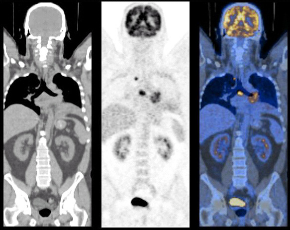
"Impetus for research" Moreover, a year from now, in addition to the cyclotron, the new Imaging Center at Hönggerberg will also have the corresponding animal PET for rats and mice and other equipment, such as an animal MRI (via the University) and optical imaging equipment. In 2007, depending on the budget, the center could also acquire the means to combine anatomical and functional analysis and examinations on mice with a CT-Animal-PET for 1.5 million CHF (see picture above). The location of the Imaging Center in the inner courtyard between the third and fourth tract of the new chemistry building at Hönggerberg makes it possible to maximise synergies between chemistry and pharmaceutical institutes. The proposed move of PSI from Villigen–in a neighbouring Canton–to Hönggerberg will mean a heavy concentration in one location of life science institutes from chemistry, physics, pharmaceuticals, biology and the materials sciences. Schubiger is confident that this will lend great impetus to research.
|
||||||||||
|
Footnotes:
You can write a feedback to this article or read the existing comments. | |||||||||||